|
In Vivo Microscopy and Mouse Imaging Live imaging technology represents a paradigm shift in how we study biological processes, from the traditional static 2D snapshots (i.e. histology) to dynamic, 3D view of the living specimen as processes unfold in real time. While whole-body imaging modalities such as MRI and CT have been highly successful in the clinical setting, mechanistic studies of biological processes such as disease progression and response to therapy in preclinical (animal) models require cellular details that are beyond the resolution of these whole-body imaging modalities. We have established a unique in vivocellular-level imaging resource to facilitate and accelerate collaborative biomedical research. This resource has strong imaging expertise as well as cutting-edge instrumentation that are developed in house and not commercially available. We develop and use confocal and multiphoton fluorescence imaging as well as other modalities. In particular, we have developed miniature high-resolution endomicroscopy that allows us to access internal organs minimally invasively in vivo and over time.
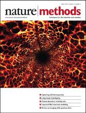
Selected papers 1. In vivo wide-area cellular imaging by side-view endomicroscopy” Nature Methods 2010 2. In vivo tracking of color coded effector, natural and induced regulatory T cells in allograft response. Nature Medicine 2010 3. Fabrication and operation of GRIN probes for in vivo fluorescence cellular imaging of internal organs in small animals. Nature Protocols 2012 4. Endoscopic time-lapse imaging of immune cells in infarcted mouse hearts. Circulation Research 2013 5. In vivo fluorescence microscopy: Lessons from observing cell behavior in their native environment. Physiology 2015
|


